CBCT
CBCT slices and volume on the patient’s own scan
The DICOM study opens from the patient record or the treatment plan: axial, coronal and sagittal slices, a panoramic along the arch curve and a 3D volume, with measurements in millimetres and captures stored with the patient.
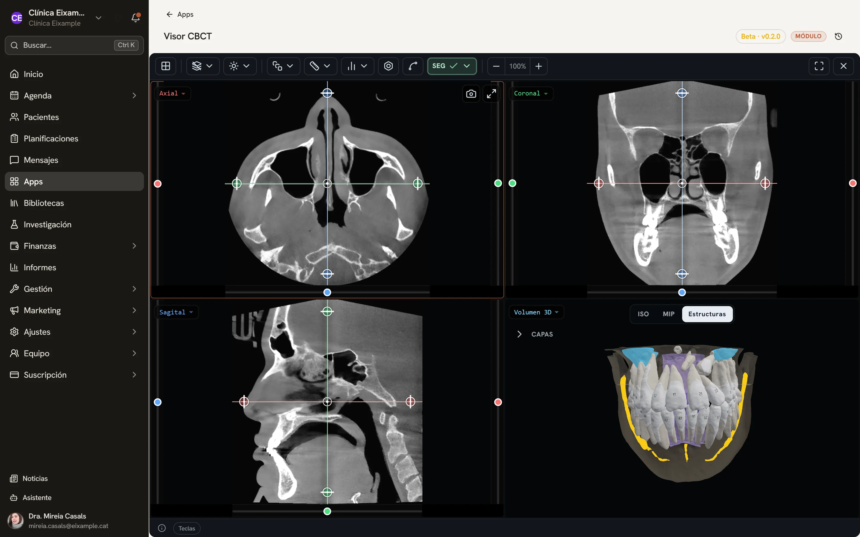
Screens of the software with sample data.
Linked slices
The three views share the same reference crosshair: moving one slice moves the others. Rotating the crosshair tilts the planes to follow the patient’s anatomy rather than the scanner table.
- Thick slab by maximum intensity (MIP) or average
- Window presets, brightness, contrast and invert
- Zoom and pan in each panel
3D volume
Volume rendering as a surface (ISO) or by maximum intensity (MIP), rotated and zoomed alongside the slices.
- Four panels at once or one in full screen
Loading the study
DICOM files (.dcm, .dicom, .ima, .img) loose or in a ZIP, dragged in or chosen from the patient record. Compressed DICOM is not supported.
Panoramic along the arch
The arch curve is adjusted on the axial slice, with its control points, position and thickness, and the panoramic is built from it.
Cross-sections
Slices perpendicular to the arch curve, laid out in a strip, to go through each area tooth by tooth.
Measurements
Distances, angles and perpendiculars on the slices, stored with the slice they were taken on. The HU value under the pointer is shown in the status bar.
Captures
The current view, as a PNG, is saved to the patient’s documents.
Permission-based access
The viewer opens with the CBCT permission, from the documents in the patient record or from the viewer step of a treatment plan.
Technical details
- Formats
- Uncompressed DICOM, loose or in ZIP
- Views
- Axial, coronal, sagittal, oblique, panoramic and 3D
- Thick slab
- MIP and average
- Captures
- PNG in the patient’s documents
- Access
- CBCT permission
See Dexodent in a demo
For a demo, a price for your clinic or any question.