Cephalometry
Cephalometric tracing with every measurement in view
Manual tracing or proposed landmarks, the classic authors’ analyses and measurements calculated from the landmarks, with their norm and deviation. Everything is stored in the patient record.
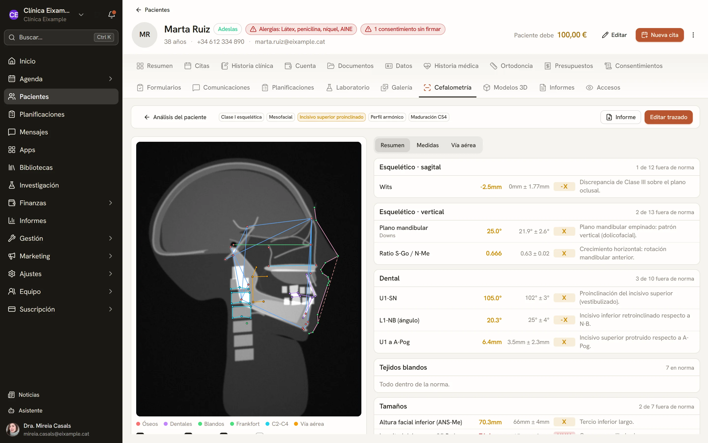
Screens of the software with sample data.
Tracing on the radiograph
Landmarks are placed in order on the cephalogram, and any of them can be repositioned with a click. With automatic detection, on the plans that include it, landmarks arrive as proposals with their confidence level and are accepted one by one or all at once.
- Cephalogram from the record or uploaded on the spot
- Calibration with the radiograph ruler
- Skeletal, dental, soft tissue and airway tracing
Summary by clinical area
Measurements are grouped into sagittal and vertical skeletal, dental, soft tissue and sizes. Each row shows the value, the norm and the deviation in X notation, and those outside the norm are flagged.
- Norms by age, sex and population group
- Full tables analysis by analysis
- Comparison between two analyses of the same patient
Analyses
Ricketts, Steiner, McNamara, Jarabak, Downs, Tweed, Holdaway and Wits, with their norm variants. Changing the analyses changes the landmarks requested and the measurements calculated.
Custom analysis
Your own analysis built from measurements in the catalogue. It is saved with the tracing and calculated like the others.
Cervical vertebral maturation
The stage is derived from the C2 to C4 landmarks with the chosen method: Baccetti (CS1–CS6), Hassel (1–6), Baccetti 2002 or Seedat.
Pharyngeal airway
McNamara upper and lower pharyngeal widths, classified with the norm by sex, plus nasopharynx, tongue base and airway angle, measured on the radiograph.
Tracing superimposition
Two tracings of the same patient superimposed on S-N at S, on the palatal plane at ANS or on the mandibular plane at Me, at the same scale when both radiographs are calibrated.
PDF report
With the chosen analyses, orientation and content, and the radiograph without landmarks, with landmarks or anonymised.
Technical details
- Images
- Lateral cephalogram in PNG, JPEG, WEBP or DICOM
- Scale
- Calibration with the radiograph ruler
- Maturation
- Baccetti, Hassel and Seedat
- Report
- Access
- Patient record and treatment plans
See Dexodent in a demo
For a demo, a price for your clinic or any question.